online advertising

Saturday, July 29, 2017

Introduction to Biochemistry - End of session 2.5 assessment - Part 3


This whole exercise is about experimental techniques - have fun!


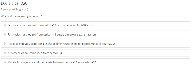
Radiolabeled fatty acids are a useful tool for researchers to dissect metabolic pathways.


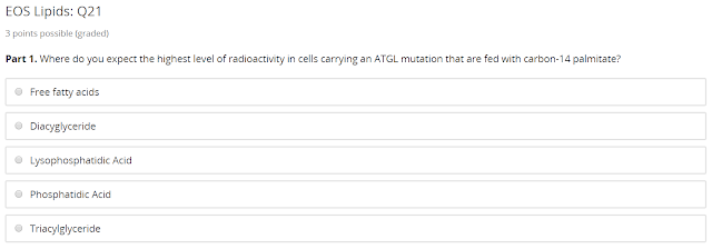
Triacylglyceride - because AGTL is not functional to turn Triacylglyceride to diacylglyceride.


Triacylglyceride - because ABHD5 cannot activate AGTL to turn Triacylglyceride to diacylglyceride.


I have a hard time to remember this one - I googled, the answer is:

Lysophosphatidic Acid - because AGPAT turns Lysophosphatidic Acid to Phosphatidic Acid.


Oleic acid is simply a fatty acid with 18 carbons, therefore the answer is:

Feed the cells with carbon-14 oleic acid and determine radioactivity in triacylglyceride relative to nonmutant cells. correct


The way to read this is that the left hand side is showing us there is a difference in these different material's polarity causing a drift in the dot. The control cell has turn all free fatty acid into triglyceride, but the mutant cell stopped at the diacylglyceride stage, which means it has problem turning diacylglyceride to triglyceride, that proves the hypothesis there is a problem with triglyceride synthesis, the answer is

My observed result matched the result predicted by my hypothesis. Therefore, the data support my hypothesis and suggest my hypothesis is correct.


As long as a wish to just click "a biochemistry college professor" or "a computer engineer who led a relatively sedentary lifestyle", the lack of physical activity has a hard time to explain why lipid is not going away in 50 years!

The only sensible option is that his body cannot get rid of lipid at all - the answer would be a patient suffering from a neutral lipid storage disease.

Introduction to Biochemistry - End of session 2.5 assessment - Part 2


A lipid droplet.


It is a mutation in a gene - therefore it is not acquired not HIV related, it is familial partial lipodystrophy.


I thought for a healthy cycle we need it to live and then die, so I chose the first one, but no, the answer is development, differentiation and survival.


If we have a genetic mutation leading to constitutive inactivation of ATGL, the lipid droplet will never be able to go through lipolysis, so lipid droplet stays.

If we have a mismatch between caloric intake and energy expenditure, lipid droplet will not be consumed fast enough and stays.

That's the two reasons why we have ectopic fat deposition.


All options involves genetic mutation in the X gene is wrong because it is mutating one gene only. Antiretroviral treatment is not a genetic mutation, so it is wrong as well. The option "Obesity due to mutations in a group of non-allelic genes" is the only correct choice.


Caloric imbalance and insulin resistance is obvious. Not sure about the chronic inflammation part at first, but it is also a correct choice. The gene mutation choices are wrong.


The guy is obviously accumulating fat. It is not a problem in triglyceride synthesis, nor it is lipodystrophy. While I guess junk food is a possible reason, you don't usually have muscle weakness.

It is likely to be an acquired metabolic syndrome, at least that's what I thought.

The correct answer is:

You suspect the patient carries a mutation in a gene regulating lipolysis and recommends a genetic test to confirm.

I would wonder, why a genetic mutation related problem shows up when the man is already 42 years old, not sure why ... there must be some environmental trigger.

Introduction to Biochemistry - End of session 2.5 assessment - Part 1


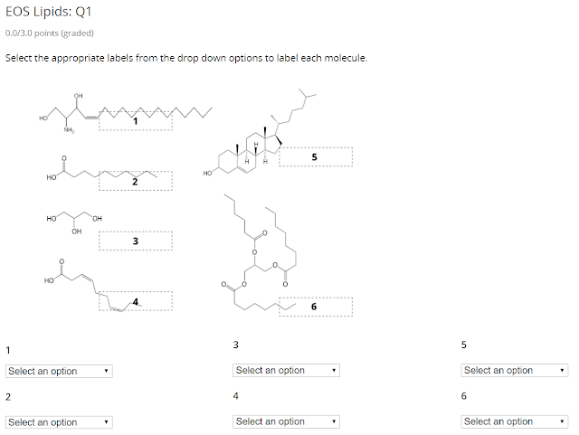
Molecule 1 is sphingosine - notice the amine group. It is a backbone molecule just like glycerol is.
Molecule 2 is a saturated fatty acid - it has a carbonxyl group and a saturated tail.
Molecule 3 is glyercol
Molecule 4 is an unsaturated fatty acid
Molecule 5 is cholesterol, with these four rings.
Molecule 6 is triacylglycerol.


Triacylglceryol is efficient energy storage, so option 1 is wrong.

While the hydrophobic repulsion of Triacylglceryol is there, that does not explain its energy density, so option 2 is wrong, same thing with option 4.

The answer is option 3, the hydration of glycogen reduces its energy density relative to triacylglycerol.


The numbering is weird, it make much more sense to name the molecules inside out.

It starts with triacylglycerol (8).
When activated, ATGL (7) can start the first lipolysis process.
That generate diacylglycerol (5)
Which is then processed by the phosphorylated HSL (6) to continue the lipolysis.
Monoacylglycerol (4) is generated in the process.
It is then processed by MGL (3) to perform the last stage of lipolysis
The fatty acid (2) that is broken down in the triacylglycerol are carried by chaperone to the blood stream because they are hydrophobic, raw exposure of fatty acid in blood stream can be toxic.
Last but not least, the glycerol (1) is also transported through the blood stream, it doesn't need a chaperone.


The answer is 4 - hormone concentration in the blood communicate the energy needs of the body to adipocytes.

After fatty meal, the blood stream has more fat than usual, there is no reason for the body to mobilize stored fats, the opposite happens. Insulin is secreted and lipogenesis occur, fats get into adipocytes. So option 1 is wrong.

Hormone circulates in blood, they are usually hydrophilic (except from steroid hormone that needs a protein carrier), and they do not bind to fatty acid directly - put it the otherway, lipolysis haven't occurred yet, where are the fatty acid to bind to? They are deep inside the lipid droplet. So option 2 is wrong.

Option 3 is almost correct, during prolonged exercise, the body experience physiological stress, the hormone epinephrine (adrenaline) is secreted by the adrenal cortex under physiological stress. The beta adrenergic receptor on the adipocyte surface will be activated to trigger the lipolysis process. This is to mobilize the energy to handle the stress.

But notice it is the adipocyte surface, not the lipid droplet surface. So option 3 is wrong, we are almost there.

Option 4 is correct, hormone concentration in blood stream is the body's way to communicate the stress to adipocyte, so that it starts to mobilize the energy.

It is nice to correlate my knowledge in human physiology with my study in biochemistry.


Glycerol is not the chaperone, so option 1 is wrong.
Chaperone shuttle hydrophobic molecules through the aqueous cytosol and blood stream, not hydrophilic, so option 2 is wrong.
Glycerol and fatty acid are useful, they are not waste, so option 3 is wrong.
The correct answer is option 4 - they are exported to liver and muscle for energy usage. It is broken down to use the energy stored in the bonds.


The outer leaflet has more cylindrical shape lipid while the inner leaflet has more conical shape leaflet, so the answer is

Sphingomyelin is most likely enriched in the outer leaflet, while
Phosphatidylethanolamine is most likely enriched in the inner leaflet.


This arrangement look so nice, it is regular packing and in paracrystaline state. This is rigid.
Adding cholesterol to it will be make it more fluid because it will no longer be so nicely packed.


This is simple, just reading graph, it is lipid 1 and lipid 2.


Conical shape things are in inner leaflet, so it is lipid 3 and lipid 4 that is conical shape.

Lipid 1 need to move from inner to outer. Floppase is needed to move lipid from inner to outer, so the answer is floppase.


I have got this wrong - I was tricked by myself about permeability by being able to let's thing in. Of course, factor that block things out are just as important - arguably more important than letting things in.

The key factor of blocking things out is the hydrophobicity. Here is the correct option:

The hydrophobic core of the lipid layer.

The key factor of lettings things in is the integral proteins. Here is the correct option:

The integral membrane proteins that allow molecule to cross.


This is simple, just reading graph, 12% is too low, low body fat is lipodystrophy.


The fill in the blanks problem is completed in the image above.

The idea is simple - lipogenesis create fat, so the lipid droplet grow, and the opposite is lipolysis. When fed, insulin store energy. So there is only one option left for the last one.


An easy one - mutation 2 causes slower growth of weight with high fat diet, the only reason is that the rat body doesn't store the intake fat, that is lipodystrophy.

Saturday, July 22, 2017

Introduction to Biochemistry - Quiz 2.5.7


In a healthy individual, fat is stored in a thin layer under the skin and around internal organs, that serves the purpose of insulation and padding.


Adipocytes contain one very large lipid droplet.


The excess fat is stored by increasing both adipocytes size and cell numbers.


Cachexia and Lipodystrophy are lack of lipid droplets, they cannot be correct because ectopic deposition is the abnormally large amount of lipid droplets.

The correct answer are the remaining two:

Metabolic syndrome resulting from caloric imbalance, and
Neutral lipid storage disease, myopathy subtype.


They are both diseases from a unique genetic mutation and Ichthyosis results from an unknown ATGL-independent function of ABHD5 that affects the skin’s lipid matrix.


It originates from dietary intake of fat, carbohydrates and proteins.

Introduction to Biochemistry - Quiz 2.5.6


12% is too few lipid droplets.


lipodystrophy and cachexia.

I think it is technically wrong reversed, I think it should be the opposite, lipodystrophy and cachexia leads to too few lipid droplets.


None of the other options make any sense, the only plausible choice is proper development, differentiation and survival.


Lipodystrophy is the loss of failure to develop adequate amounts of adipose tissue for energy storage.


This is from my memory in the lecture, it is mutation in the AGPAT.


This is also from my memory in the lecture, it is AKT2 and nuclear receptor PPARy.

Introduction to Biochemistry - Quiz 2.5.5


It is the fluorescently-labeled lipids.


At $ t = 0 $, the laser has bleached the lipid dye at this spot.

At $ t = 100 $, the lateral diffusion of the bleached and fluorescent lipids caused the dark spot to be mixed in.


Membrane protein serves their functions, so depending of the function of the cell or organelle, the percent mass of lipid versus protein will vary.


Red blood cell travels around the body, it is easy to imagine it need to perform many different functions, and therefore it has a lot of different proteins on its membrane. Photoreceptor, however, serve one purpose, to sense light, so it is reasonable to predict there is fewer types of protein on it.

Here is the answer.

The protein composition of a membrane reflects its function.

For example, while red blood cells and photoreceptors both have a high density of protein in their plasma membrane, red blood cells have a greater variety of protein while photoreceptor cells have essentially one type of protein in their membranes.


Basically integral protein can go through the membrane while peripheral protein are on one side, either outside or inside, so it is not hard to figure out:

Integral membrane protein must have hydrophobic coils that pass through the core of the lipid bilayer.
Peripheral membrane protein can be covalently linked to a lipid that is inserted in the lipid bilayer
Integral membrane protein can transduce an extracellular signal across the lipid bilayer.
Both membrane protein can serve functions on the intracellular and extracellular surfaces of the lipid bilayer.

The last one is tricky, I was tricked.


The first one is correct, water is very concentrated in the body - and the high concentration make a high concentration gradient if diffusion is blocked.

Many molecule are polar and can hydrogen bond, even a whole protein molecule can, this is wrong, being polar and can hydrogen bond alone cannot lead to diffusion.

Water molecule is small, and small molecule can easily go around barrier, this is correct.

While it is true that water is needed in the cytosol (for example, for protein folding), this is not a reason, so this is wrong.

Water is not hydrophobic and it is not bulky at all, so this is wrong.


The concentration gradient for these ions are crucial for neural signalling, they are maintained through pumps. These pumps are integral membrane proteins, so the answer is.

The ions require integral membrane proteins to cross.

Thursday, July 20, 2017

Introduction to Biochemistry - Quiz 2.5.4


Of the phospholipid shown above, those with a cylindrical shape are more likely to be found in the outer leaflet of the membrance, while lipid with a conical shape are more likely to be found in the inner leaflet.


Scrambase moves lipids down their concentration gradient, in either direction.
Floppase moves lipids against their concentration gradient, from inner to outer membrane.
Flippase moves lipids against their concentration gradient, from outer to inner membrane.
Scrambase actively promotes membrane compositional symmetry.


In the endoplasmic reticulum.


Sphingomeylin is synthezised in the lumen of the golgi apparatus and is transported to the plasma membrane by vesicles Sphingomeylin will first be incorporated into the outer leaflet of the plasma membrane.

This is because the inside of the vesicle contains the material that the cell wants to secret, so that will face outside after the vesicle merge with the plasma membrane.